04.01 / FRACTURES & TRAUMA
Wrist
Fractures
The same fall can produce very different wrist injuries. Understanding which bone is involved, how it is aligned and whether it is stable helps determine the right protection, treatment and route back to activity.
01 / THE WRIST END OF THE FOREARMAlignment & joint surface
Distal Radius
Fracture
What it is
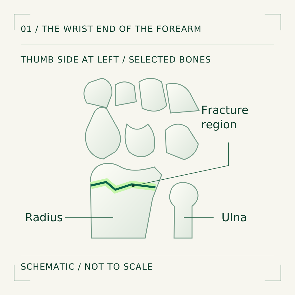
The radius is the thumb-side forearm bone. A distal radius fracture is a break near its wrist end. Fragments may remain aligned, shift or involve the joint surface. Nearby structures can also be injured.
1What you may notice after the fall
Wrist pain, swelling and bruising are common, with difficulty moving or using the hand. A displaced fracture may change the wrist’s shape. A wrist can still be fractured without a visible deformity, so persistent pain after injury needs assessment.
1A fall, a collision or a higher-energy impact
Landing on an outstretched hand is a common mechanism. Sport, falls from height and road collisions can transmit greater force and produce more complex injuries. The mechanism helps the team consider damage beyond the bone as well as the fracture itself.
3Bone strength changes the force needed
People of different ages can sustain this fracture. Fragile bone may break after a relatively minor fall; stronger bone can break during a substantial impact. A low-energy fracture in an older adult can prompt discussion of bone health and ways to reduce future falls.
2Position now, and the ability to hold it
Displaced fragments have shifted. An unstable fracture risks losing an acceptable position, including after realignment. Fragmentation and joint involvement help determine treatment and follow-up.
1Understanding the problem.
Choosing the next step.
Checking the wrist and the whole hand
Assessment records how the injury happened and checks skin, swelling, finger movement, sensation and blood flow. The clinician also considers associated injuries and your usual work or activity. Significant deformity or nerve symptoms may require prompt treatment before a longer-term plan is agreed.
2X-rays first; more detail when it changes care
X-rays show the break, alignment and whether the wrist joint is involved. A CT scan may clarify a complex joint-surface fracture or help plan fixation. Follow-up X-rays can check that a fracture remains in position during early healing.
1Protection, and reduction when needed
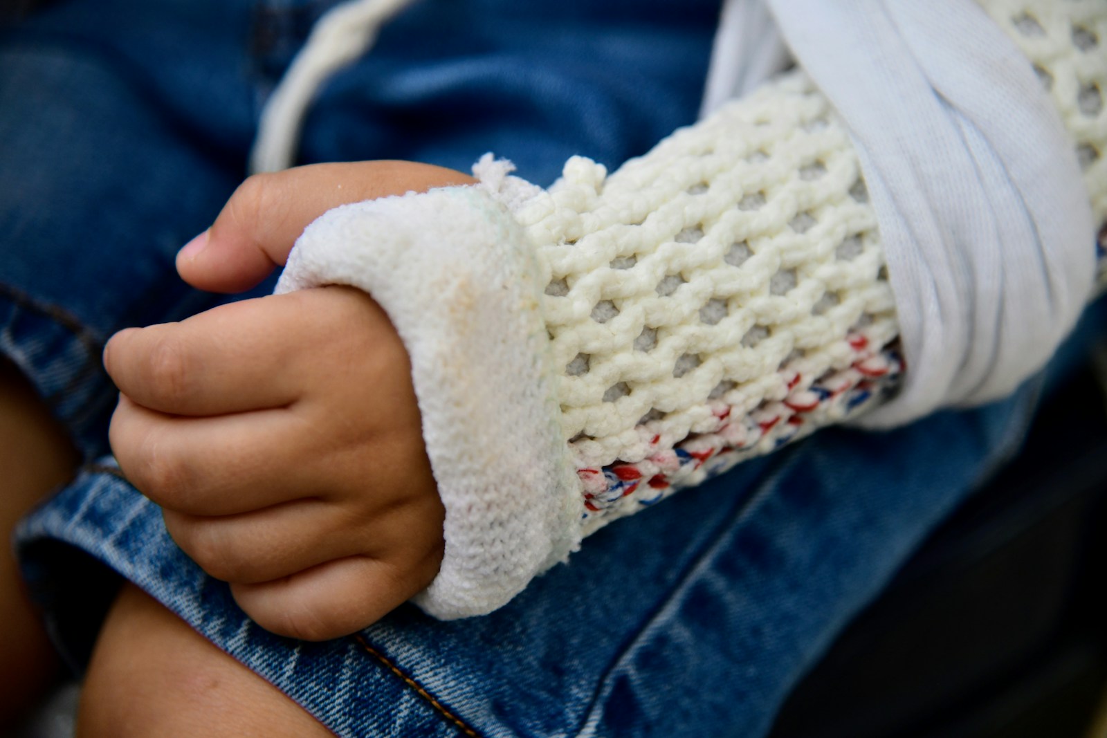
A suitably aligned fracture may be protected in a splint or cast. If the fragments have shifted, a clinician may realign them under appropriate pain control or anaesthesia; this is called reduction. The wrist then needs protection and review to check that the position is maintained.
3Holding the fragments in a useful position
Fixation may be considered when acceptable alignment cannot be achieved or maintained, or the joint surface needs restoration. Plates and screws, wires or external fixation are selected according to the injury. The potential functional benefit is weighed against surgical risks and the individual’s priorities.
2Protection and movement need to work together
Keep the cast or splint as instructed and report pressure or increasing discomfort. Swelling control and prescribed movement of joints that are safe to move help limit stiffness. Wrist exercises begin when the treating team allows; removing protection early can put healing at risk.
3Healing is followed by rebuilding function
Stiffness, swelling and reduced grip can continue after the cast is removed or the fracture has united. Hand therapy may guide mobility and gradual strengthening. Light activities generally precede heavy loading. Return to work or sport depends on healing, movement, strength and the demands of the task, with progress varying between people.
4When assessment is urgent
A deformed wrist, a wound, new numbness, pale or cold fingers, or severe increasing pain needs urgent care. The same applies if symptoms develop in a cast. Do not wait for a routine appointment when circulation or nerve function may be affected.
102 / THE THUMB SIDE OF THE WRISTA small bone with a distinct healing pattern
Scaphoid
Fracture
What it is
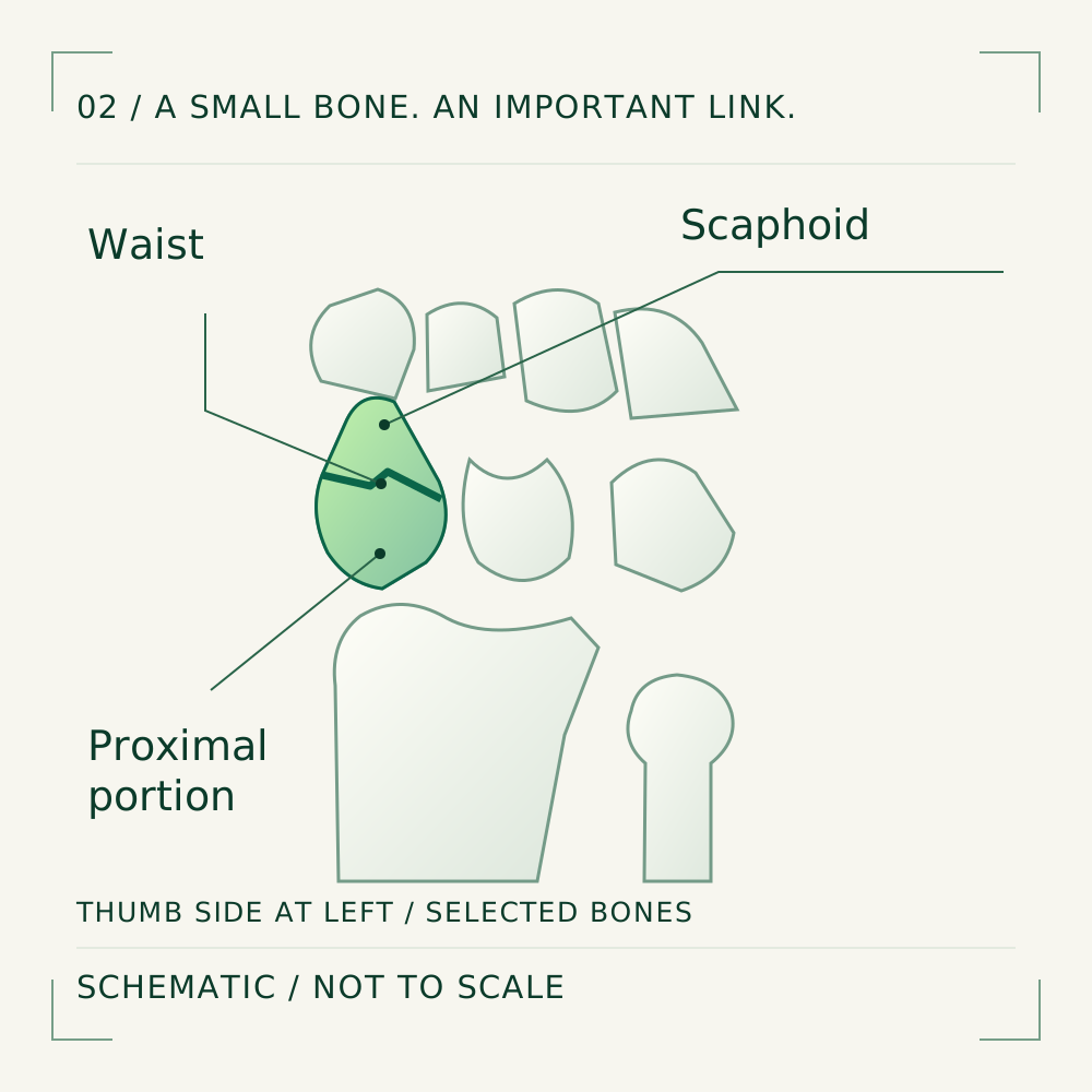
The scaphoid is one of the small wrist bones, linking the two carpal rows on the thumb side. A fracture can occur through its middle, called the waist, or nearer either end. Its location and blood supply influence healing and the choice of treatment.
5A subtle injury can still matter
Pain is often on the thumb side, including the hollow behind the thumb tendons known as the anatomical snuffbox. Gripping, pinching or loading the wrist may hurt. The absence of obvious deformity does not establish that the injury is only a sprain.
7Force through an extended wrist
A fall onto the palm can load the scaphoid while the wrist is bent backwards. Sports injuries and road collisions are other mechanisms. The same incident can damage additional wrist structures; the painful spot is a starting point for examination, not the whole diagnosis.
5Everyday falls as well as sport
Scaphoid fractures occur across age groups. Activities that risk a fall onto an outstretched hand are relevant, but a dramatic accident is not required before thumb-side wrist pain deserves attention. Describe the position of the wrist and what happened immediately after the injury.
5Why the site of the break affects healing
Some blood vessels enter nearer the thumb end and supply the portion closer to the forearm. A fracture can interrupt that supply. Delayed union means slower healing; non-union means the fracture has failed to unite. Reduced blood supply can also damage bone, sometimes called avascular necrosis.
5Understanding the problem.
Choosing the next step.
Examine the painful area and arrange follow-up
The clinician checks tenderness over the scaphoid, swelling and wrist function, and reviews the mechanism. Examination alone cannot reliably exclude a fracture. If it remains suspected, a clear plan for protection, imaging and review is needed rather than simply waiting for the pain to disappear.
6Different scans answer different questions
Initial wrist X-rays include views intended to show the scaphoid. MRI can identify an injury missed on early films; CT helps define the fracture and alignment. Repeat examination and X-rays may be used in some pathways. The choice depends on findings, timing and local access.
7A cast can be appropriate for a stable fracture
Many non-displaced fractures can be managed with immobilisation. The cast or splint and duration are chosen for the fracture site and progress. Protection is still important while a suspected injury is being investigated. Follow the specific instructions given rather than testing the wrist by repeatedly loading it.
6Fixation when the pattern or healing requires it
A displaced or selected higher-risk fracture may need fixation, often with a screw. Established non-union can require bone grafting as well as stabilisation. Surgery aims to support union and useful alignment; it does not guarantee healing or remove the need for protection and follow-up.
7Keep the rest of the arm moving safely
While following the prescribed wrist protection, gentle movement of permitted fingers, elbow and shoulder can reduce stiffness. Avoid heavy lifting or gripping until cleared. Ask which light tasks are safe for your particular injury; advice for an undiagnosed wrist injury may change once a fracture is confirmed.
8Symptoms alone do not prove union
Healing can take longer than expected, and a quieter wrist is not by itself proof that the bone has joined. Review and imaging guide progression. Work and sport resume according to union, safe function and loading demands; contact sport and fall-risk activities may need a longer period of protection.
7Early review prevents a missed injury being overlooked
Seek prompt assessment for persistent thumb-side pain after a fall, even after a reassuring first X-ray. Attend the planned scan and results review, and contact the treating team if these have not been arranged or symptoms worsen. Early diagnosis gives the fracture a clearer treatment pathway.
8THE NEXT CHAPTER / YOUR CARE
After an injury, alignment and function both matter.
Specialist assessment can clarify the fracture pattern, stability and next steps for recovery. A deformed wrist, worsening severe pain or changes in finger sensation or circulation need urgent care.
Book a Consultation